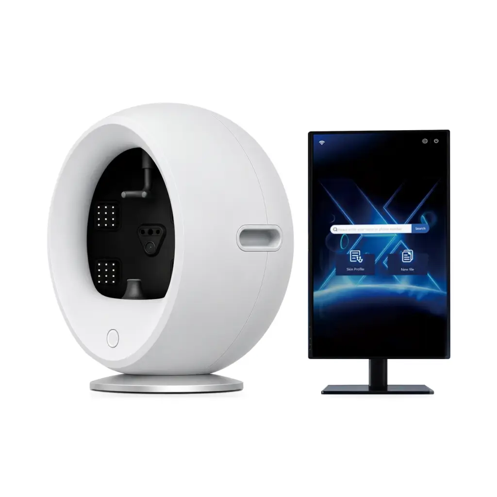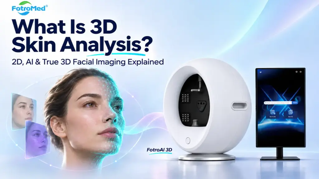

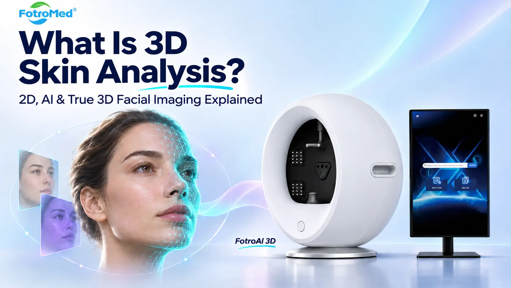
True 3D skin analysis combines two technologies in a single scan:
1. Multi-light skin imaging to look beneath the skin’s surface at pigment, Rötung, Poren, und UV -Schaden.
2. True 3D facial reconstruction to capture the real shape, Tiefe, and contours of the entire face.
When combined, practitioners can inspect skin quality Und rotate a lifelike 3D digital model of the patient’s face to check cheek volume, Kieferdefinition, and facial symmetry.
If you are shopping for a skin analysis machine for your med spa or clinic, you have probably noticed a confusing trend. Almost every device on the market claims to offer „3D skin analysis“ oder „AI skin imaging.“ Yet prices range from $500 tablet attachments to $30,000 clinical booths. Why is there such a massive gap?
The short answer is simple: most „3D“ skin analyzers on the market do not actually capture 3D faces. Many devices simply add digital shading to flat 2D photos, while others rely on basic tablet cameras paired with AI apps. Owners of high-end aesthetic centers, dermatologists who offer fillers, and injectors focused on facial contouring need imaging that clearly documents changes in facial volume and contour before and after treatment. A system built for selling facial creams will not help you plan dermal fillers, HIFU lifting, or laser resurfacing. In diesem Ratgeber, we will break down what 3D skin analysis really means, explain the exact differences between 2D-Bildgebung, AI imaging, and True 3D facial imaging, and show you how to choose the right machine for your practice.
What Is 3D Skin Analysis? (And the „Fake 3D“ Trap)
Watch Out for „Pseudo-3D“ (Fake 3D)
Vor dem Maschinenvergleich, buyers need to understand a common marketing trick known as pseudo-3D.
Many budget 2D analyzers advertise „3D wrinkle and pore views.“ Here is how they actually work: the machine takes a flat 2D photo and measures how light or dark each pixel is. The software assumes dark pixels are deep valleys and bright pixels are high peaks, turning them into a colorful bumpy chart.
While this trick can help visualize rough skin texture on a small patch of skin, it is not true 3D facial imaging:
- If a natural shadow falls across the nose or cheek, the software mistakes that shadow for a deep hollow.
- It cannot measure real facial contours, chin projection, or midface volume.
- You cannot rotate the full face to show a patient their side profile or oblique angles.
2D vs. AI Imaging vs. True 3D Facial Imaging: What Is the Difference?
People often talk about 2D, Ai, and 3D imaging as if they are three competing types of cameras. In Wirklichkeit, 2D and 3D describe how the hardware captures the image, während AI describes how the software reads that image.
Let’s look at what each technology actually does in a consultation room.
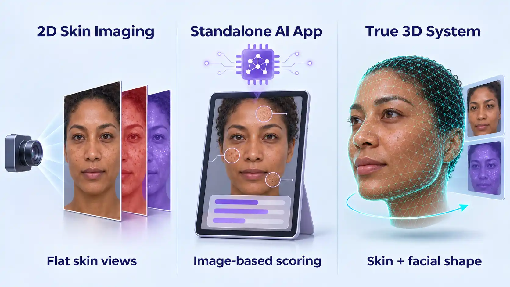
1. 2D Multi-Spectral Skin Imaging: Great for Color, Blind to Shape
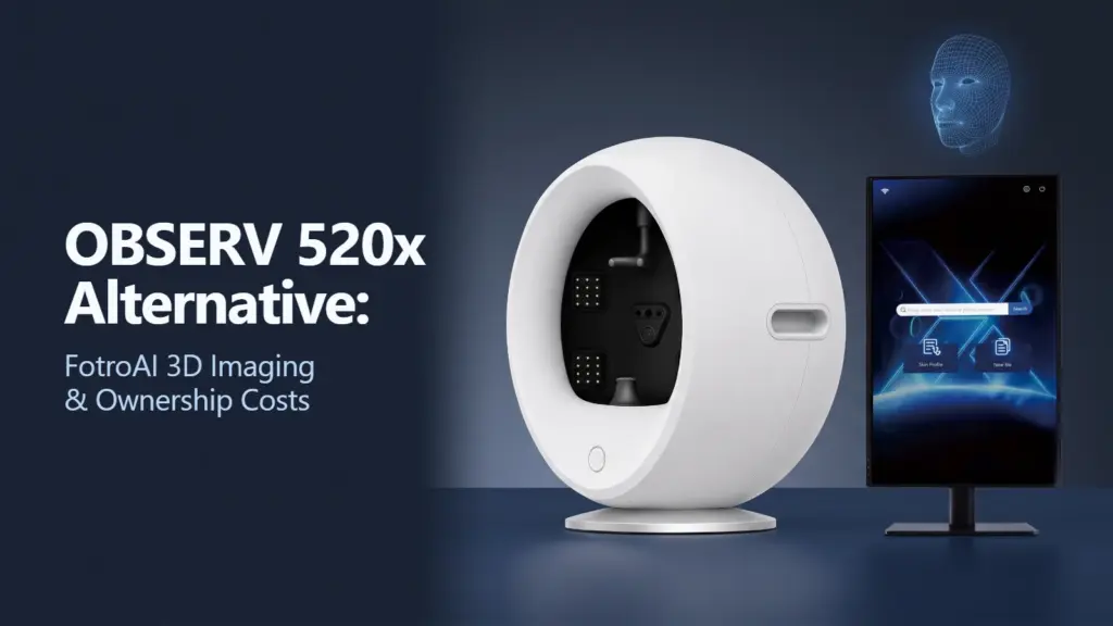
Traditional 2D skin analyzers use a single camera paired with specialized lighting—typically standard white light (RGB), polarisiertes Licht, und UV-Licht. Portable tablet-based hoods, such as the Sylton Observ series, are popular examples of multi-light 2D systems.
- Wie es funktioniert: Special light filters cut through surface glare to reveal hidden brown spots, broken capillaries, verstopfte Poren, und Sonnenschäden.
- Where it shines: It is reliable and effective for basic dermatology, acne facials, chemische Peelings, and pigment treatments.
- Where it falls short: Everything stays flat. A standard 2D photo cannot directly measure facial depth or volume. If you perform Botox, Füllstoffe, Fadenlifte, RF-Mikronadelung, oder Hifu, subtle changes in contour can be hard to assess from flat images, especially when head position changes between visits.
Clinical measurements from 2D photos and structured-light scans
Cascos and colleagues used direct caliper measurements as the reference. They compared distances between facial landmarks from 2D photos and structured-light scans in 60 Teilnehmer. With the teeth together, the average differences from the reference were 2.09 mm for 2D photos and 0.61 mm for structured light. During a smile, they were 2.49 mm Und 0.28 mm, jeweils. The figures apply to the equipment and measurements tested.
Cascos et al., 2023 facial measurement study
2. AI Skin Imaging: Der „Brain,“ Not the „Augen“
AI skin imaging uses computer vision to automatically detect skin concerns, count spots or fine lines, calculate a „Hautalter,“ and recommend treatments.
- Wie es funktioniert: The software applies patterns it learned during training to a new photo and returns automated scores in seconds.
- Where it shines: Speed and consistency. Entry-level AI apps running on iPads or smartphones are cheap, schnell, and easy to use at retail skincare counters.
Image quality affects AI skin scores.
Lee and colleagues enrolled 1,099 participants in South Korea. They trained and tested separate AI models on DSLR, Tablette, and smartphone photos, using dermatologist grades as the reference. For forehead pigmentation, the AI grade matched the dermatologist grade exactly in 58.7% of DSLR cases, 55.2% of tablet cases, Und 50.5% of smartphone cases. The figures reflect the tested devices and this specific skin feature.
Lee et al., 2026 AI skin assessment study, Tisch 9
3. True 3D Facial Imaging: Built for Contours, Heben, and Precision
True 3D facial imaging captures real physical geometry—width (X), height (Y), and actual depth (Z).
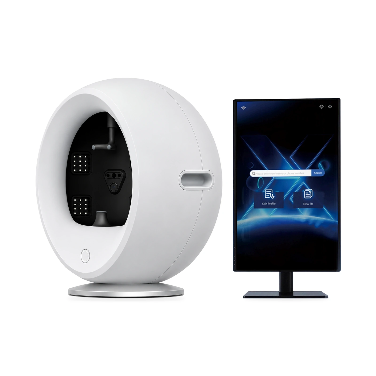
- Wie es funktioniert: Structured-light scanning projects a known pattern onto the face and uses its distortion to measure depth. Multi-camera stereophotogrammetry calculates depth from images taken at different angles. The active structured-light system gave the measurements closest to the manual reference and the most consistent repeat results among the four facial scanning systems tested in 2026. Fotromed specifies that the FotroAI 3D combines structured light and three 20MP cameras to build a 45-megapixel full-face model with 0.2 mm reconstruction accuracy.
- Where it shines: Practitioners can smoothly move between frontal, oblique (45-Grad), and profile views within one digital model. You can clearly assess facial proportions, asymmetry, cheek contours, chin projection, and lower-face laxity. When paired with multi-spectral lights, you get deep skin diagnostics Plus exact facial structure in one quick scan.
- Where it falls short: Because it requires precision optical sensors and controlled lighting, it is a dedicated desktop console rather than a pocket device. Jedoch, modern manufacturing has made true 3D consoles far faster and much more affordable than older hospital systems.
💡 Optical Lab & Clinic Field Note:
In our optical calibration tests at FotroMed, a chin tilt of just 5 degrees upward on a standard 2D analyzer makes lower-face laxity and submental shadows appear noticeably tighter—creating a „false positive“ lifting result at follow-up visits. Likewise, unshielded warm ceiling spotlights in a treatment room can skew tablet-based redness scores. Using an enclosed multi-light chamber and locking 3D facial geometry solves both issues at the source.
Nebenseite Vergleich: 2D vs. AI Apps vs. True 3D Systems
| Besonderheit | 2D Multi-Spectral Analyzer | Standalone AI App (Phone/Tablet) | True 3D + Multi-Light System |
|---|---|---|---|
| Core Hardware | Single camera + filtered lights | Tablet or smartphone camera | Multi-camera array + structured light + multi-spectral LEDs |
| Captures Real Facial Shape? | NEIN (flat images or „pseudo-3D“ shading) | NEIN | Ja (sub-millimeter 3D geometry, z.B., 0.2 mm-Genauigkeit) |
| Sees Subsurface Pigment & Rötung? | Ja (via polarized & UV -Licht) | Beschränkt (surface RGB only) | Ja (RGB, parallel/cross-polarized & UV views) |
| Multi-Angle Contour Review? | Requires separate manual photos | NEIN | Ja (smooth rotation across front, oblique & profile) |
| Vor & After Reliability | Good for color; sensitive to head tilt | Niedrig (affected by room light) | Hoch (3D angle alignment + color calibration) |
| Am besten für | Facials, schält, basic pigment care | Skincare retail, online quizzes | Injektionspräparate, HIFU/RF lifting, Laser, plastische Chirurgie & mit Spas |
Why Leading Clinics Combine True 3D, Multi-Light, and AI
Modern aesthetic clinics rarely stick to just one type of treatment. A single patient might come in for IPL or a laser to clear brown spots, RF tightening for their jawline, and dermal fillers for midface volume.
VISIA and ANTERA 3D produced correlated skin texture measurements. Ihre pore and wrinkle measurements showed no linear correlation. That study examined small skin areas. A full-face 3D scan addresses facial contours across a much larger area. Because skin quality and facial structure go hand in hand, equipment makers combine True 3D modeling, multi-spectral lighting, and smart software into one platform.
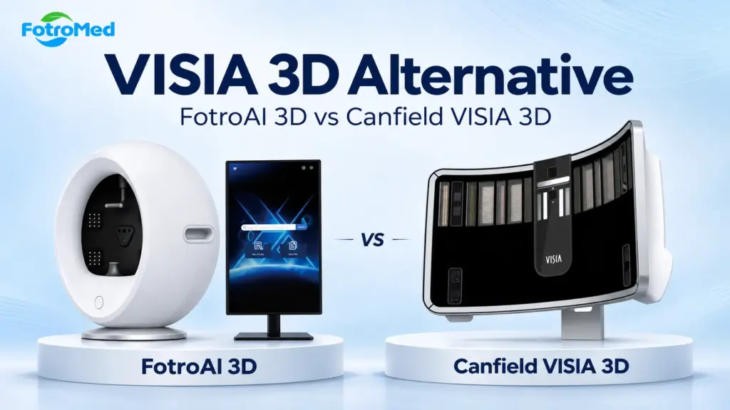
Following this exact clinical standard, next-generation systems like FotroMed’s FotroAI 3D bring clinical-grade 3D reconstruction and multi-light skin diagnostics together—without the steep price tag or ongoing software fees often tied to legacy booths. If you are comparing specific models, Merkmale, and clinic budgets against legacy systems, read our complete guide to the top alternatives to the VISIA skin analysis machine.
Which Technology Fits Your Aesthetic Business?
Das Recht wählen Hautanalysemaschine comes down to your treatment menu:
- Choose a 2D + AI Tablet Tool if: You run a cosmetic retail counter, beauty bar, or online skincare brand where quick, low-cost product recommendations matter more than clinical documentation.
- Choose a 2D Multi-Spectral Analyzer if: Your studio focuses strictly on surface facials, Akne -Pflege, and basic pigment treatments, and you do not offer contouring, Straffung, or injectables.
- Choose True 3D Facial Imaging if: You operate a mit Spa, dermatology clinic, injectable practice, or plastic surgery center and need to track changes in facial shape or volume. Researchers used 3D facial images to measure changes in chin volume after hyaluronic acid filler at follow-up visits. A baseline scan and matching follow-up views help clinicians show patients where their contours changed. Multi-light views add a record of skin appearance for clinics that also provide skin treatments.
For a model-by-model comparison, siehe unsere bestes Hautanalysegerät roundup.
Upgrade Your Consultations with True 3D Clarity
Patients invest in aesthetic treatments when they can clearly see what their skin needs—and see proof that your treatments work. Moving beyond flat 2D photos and „pseudo-3D“ gimmicks gives your practitioners the visual authority to plan comprehensive, high-value treatment packages with confidence.
Häufig gestellte Fragen (FAQs)
1. Was ist der Unterschied zwischen „pseudo-3D“ and true 3D skin analysis?
Pseudo-3D takes a flat 2D photo and uses light and dark pixels to guess skin texture, meaning natural facial shadows can easily be mistaken for deep hollows. True 3D facial imaging uses structured light and multiple cameras to measure real physical width, height, and depth—creating an accurate 3D face model you can rotate to examine real contours.
2. Can a regular 2D skin analyzer measure facial contours for fillers or HIFU?
NEIN. Standard 2D analyzers only capture flat images. While they can highlight spots and pores, they cannot accurately show cheek projection, facial asymmetry, or jawline lifting from multiple angles on a unified 3D model.
3. Is AI skin analysis the same as 3D skin analysis?
NEIN. AI refers to the software that scores and analyzes an image, while 3D refers to optical hardware (such as structured light and multi-camera arrays) that measures real physical depth and facial shape. The best modern clinical machines run smart analysis software on top of true 3D and multi-spectral hardware.
4. How accurate is true 3D facial imaging, and how long does a scan take?
The FotroAI 3D specifications list a 45-megapixel full-face model, 0.2 mm reconstruction accuracy, und a 20-second full-face scan. Those are Fotromed’s figures for this model.
5. Why does a 3D skin analysis machine still need polarized and UV light?
True 3D scanning maps facial shape, Symmetrie, and contours. Parallel-polarized light brings out surface detail. Cross-polarized light reduces surface glare so color variations such as redness are easier to see. UV imaging can reveal patterns associated with sun damage or skin fluorescence such as porphyrins.
6. Is structured-light 3D facial scanning safe for all patients?
Structured-light 3D capture uses a projected light pattern and works without touching the skin. The IEC 62471 standard recognized by the FDA describes how to evaluate optical radiation from lamps, including LEDs. Check the device instructions for each imaging mode, including any eye protection and restrictions on use. Separat, the 48-color ColorChecker helps clinics keep image color consistent between visits.
7. Do all professional 3D skin analysis machines charge annual software fees or require consumables?
Not all of them. While several legacy brands charge ongoing software licensing, Wolke, or support fees, systems like the FotroAI 3D require no disposable consumables and include full 3D imaging, 8-view multi-light analysis, and multi-location patient management software with zero subscription or renewal fees.
Editorial disclosure. Fotromed offers the FotroAI 3D featured in this guide. This article helps aesthetic practitioners and clinic operators compare imaging equipment. Optical images and 3D models support consultation, Planung, und Dokumentation. Clinical and histopathological diagnoses are the responsibility of qualified healthcare professionals.
Referenzen & Quellen
- Cascos R et al. Accuracy between 2D Photography and Dual-Structured Light 3D Facial Scanner for Facial Anthropometry: A Clinical Study. Journal of Clinical Medicine, April 2023.
- Hanlon KL et al. Dermoscopy and skin imaging light sources: a comparison and review of spectral power distribution and color consistency. Journal of Biomedical Optics, August 2022.
- Burlacu Vatamanu OE et al. Evaluation of Four 3D Facial Scanning Technologies: From Photogrammetry to Structured-Light Systems in Clinical Dentistry. Dentistry Journal, Februar 2026.
- Marcum KK, Goldman ND, Sandoval LF. Comparison of standard, parallelpolarisiert, kreuzpolarisiert, and ultraviolet skin photography. Zeitschrift für Arzneimittel in der Dermatologie, Februar 2015.
- Jung G, Yoo S. Ultraviolet-induced fluorescence mapping of facial porphyrin and sebum using deep-learning segmentation. Photodiagnosis and Photodynamic Therapy, December 2025.
- Lee E et al. Artificial Intelligence Based Skin Analysis Models for Predicting Visual Grades and Device Measured Physiological Values From Facial Images. Skin Research and Technology, September 2026.
- Sylton. OBSERV 520x official product overview, positioning and light modes.
- Canfield Scientific. Canfield Care annual support and software upgrade agreements.
- Voytik M et al. Three-Dimensional Volumetric Analysis of Chin Augmentation Using Hyaluronic Acid Filler: A Prospective Study. Plastic and Reconstructive Surgery, online July 22, 2026.
- Spanish Society of Aesthetic Medicine. Comparison of aesthetic imaging systems and European equipment price estimates, 2026.
- UNS. FDA. Recognized standard IEC 62471:2006 for photobiological safety assessment of lamps and lamp systems.